. DOI: 10.1016/J.NEUROIMAGE.2025.121063")
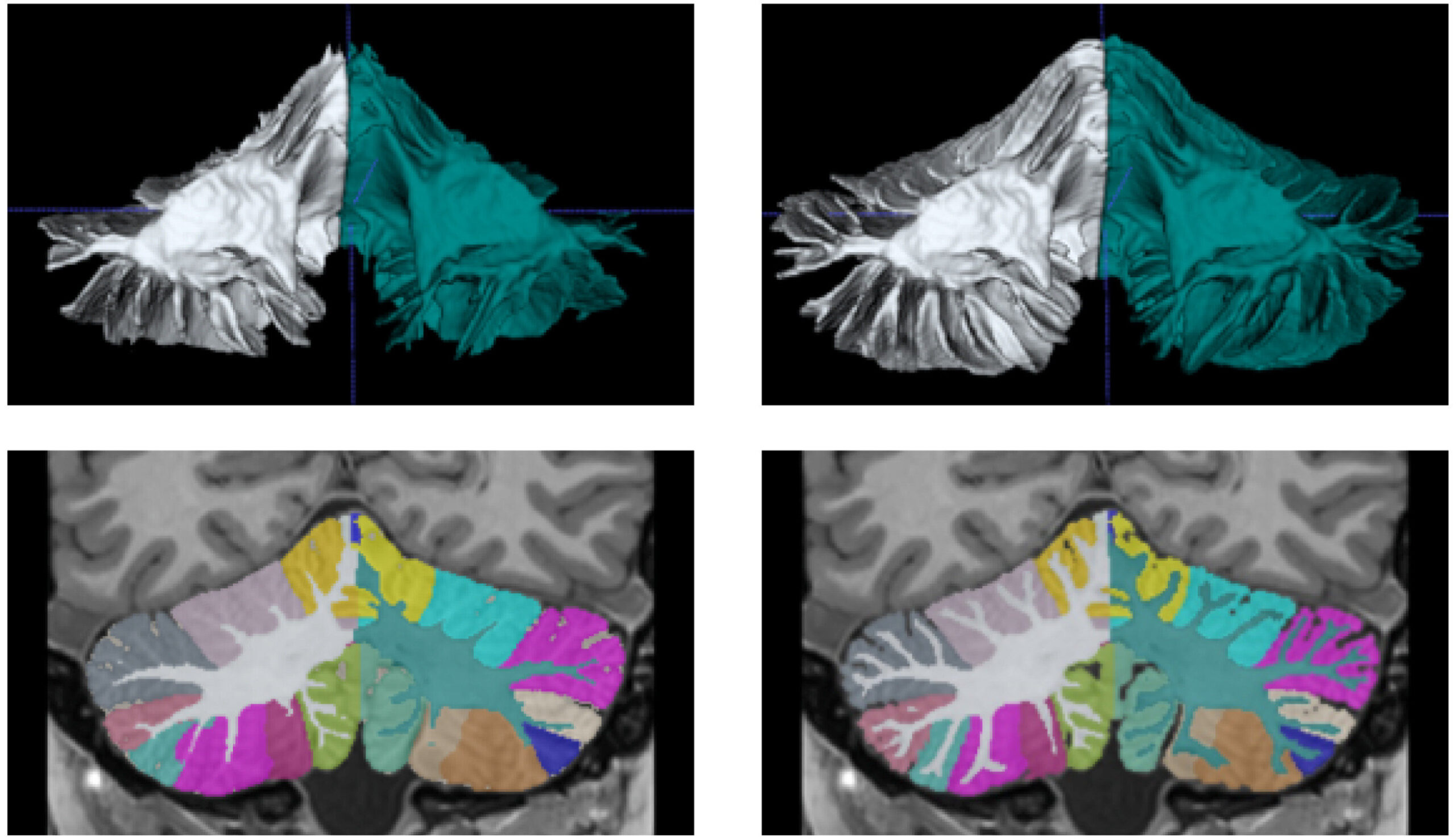
Left: 3D Reconstruction of Wm Label and A Coronal View of Segmentation Before Semiautomatic Correction. Right: 3D Reconstruction of Wm Label and a Coronal Segmentation View after Semiautomatic Correction. The Wm “Arbor Vitae” is Better Defined in the Corrected Version. CREDIT: Neuroimage (2025). DOI: 10.1016/J.NEUROIMAGE.2025.121063
Team of Researchers from the Universitat Polytècnica de València (UPV) and the French National Center for Scientific Research (CNRS) has developed the World’s Most Advanced Software to Study The Humabellum Using High-Resolution nmr Images.
CALLED DEEPCERES, this software will hell HELP in the Research and Diagnosis of Diseases Such As als, Schizophrenia, Autism and Alzheimer’s, Among Others. The Work of the Spanish and French Researchers has Been Published in the Journal Neuroimage.
Despite its Small Size Compred to the Rest of the Brain, The Cerebellum Contains Approximately 50% of All Brain Neurons and Plays The Fundamental Role In Cognitive, Emotional and Motor Functions.
As Sergio Morell-Ortega, Project Research at the Itaca Institute of the Universitat Polytècnica de València, Explains, Segmentation of the Cebellum Has Until Now Been A Great Challenge Due to the Complexity of It Anatomy and the Difficulty Of Differentiating Its Structures by Means of Conventional Magnetic Resonance Imaging.
“Deepceres overcomes all these challenges and is, today, the mos accurate tool in the world for measuring such an important structure of the central nervids System as the cerebellum,” emphasizes more.
High Accuracy
The Deepceres Software is Capable of Measuring 27 Structures of the Cerebellum. And stands out above all for improving the precision of segmentation compredo to what is ahieved with the methods use to date, Thanks mainly to the application artificial artificial intelligence tools.
“Using Standard Resonance Images of 1 Cubic Millimeter, these are converted into ultra-high resolution images of 0.125 mm3 Using Deep Neural Networks, “ADDS Professor José Vicente Manjón, The Main Researcher of the Project.
“This AllWs Researchers and Health Care Professionals to ObtaIn Detailed Information About the Anatomy of the Cerebellum Without The Need For Ultra-High-Resolution Date in the Initial Image. It’s Like Going From A Black-and-White Image to Color Image. and, Moreover, It is Accessible to the Entire Scientific Community. “
Applications in Neuroscience and Clinical Practice
ACCORDING TO THE DEVELOPERS OF DEEPCERES, THE PRECISION IN THE VOLUMER QUANTIFICATION OF THE CERELLUM WILP IN THE STUDY OF NEUROLOLOGICAL PATHOLOGIES SUCHO AMYOTROPHIC SIDE SLECTROSIS OR PSYCHIATRIC ILLNESSES SUCHO AS SCHIZOPHRENIA AND AUTISMM.
“FURTHERMORE, DIFFERENT STUDIES PUBLISHED RECENTLY HAVE DEMONSTED THE INCIDENCE OF THE STRUTURE OF THE CERBELLUM IN NEURODEGegenerative Diseases Such As Alzheimer’s,” ADDS SERGIO MORELL.
15,000 cerebellums in five months
To Facilitate its use, the UPV and French CNRS Teams have developed an Online Platform That is Accessible to Research and Medical Staff. Since its Launch Just Five Months ago, Deepceres has processed images of Nearly 15,000 cerebellums. To date, it has been used by experts from many country, with the greatest impact in the United States and China.
Researchers from the Research Institute of Industrial Control Systems and Computing and the Applied Mathematics Department Polytècnica de València, The Department of Psychobiology at the University of Valencia, The Medical Imaging Department at La Fe University and Polytechnic Hospital and the fisabio prince Felipe Biomedical Research Center joint biomedical imaging unit have also participated in its develoopment.
More information:
Sergio Morell-Ortega et al, Deepceres: Deep Learning Method for Cerebellar Lobule Segmentation Ultra-High Resolution Multimodal MRI, Neuroimage (2025). DOI: 10.1016/J.NEUROIMAGE.2025.121063
Citation: Deepceres: Ai-Driven Software Redefines Cebellum Research with Detailed Imaging (2025, March 20) Retrieved 20 March 2025 from
This document is Subject to Copyright. Apart from Any Fair Dealing for the Purpose of Private Study or Research at Part May Be Reproduced Without The Written Permission. The Content is Provided for Information Purposes Only.
